How would you report this ECG?

For those unfamiliar with ECGs, let us look at how a 12-lead ECG is generated.
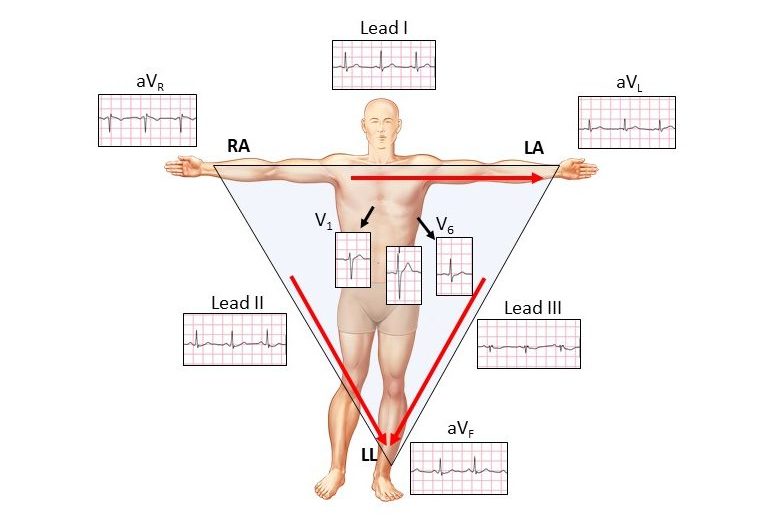
Einthoven’s triangle is formed by three skin electrodes; right arm (RA), left arm (LA) and left leg (LL). It is an inverted equilateral triangle with the heart at its centre.
Lead I is created between the negative potential RA electrode and the positive LA electrode and is usually positive as the current flows from R to L (red arrow). For leads II and III, the left leg is positive. The right leg electrode by convention is the earth. The three unipolar augmented leads (aV) are also dependent on the polarity with aVR negative. The chest leads V1 to V6 traverse the chest wall.
We can now create a normal 12-Lead ECG:

Knowing this, our first ECG looked different because of reversed limb leads.
For the example shown, the arm and leg leads are reversed.

When this occurs, lead I (blue arrow) is now on the legs and is therefore isoelectric (red highlight). This is a major footprint which should immediately catch the eye. The other changes are minor footprints and are not always reliable, apart from lead aVL being flipped (yellow highlight), as that lead is now on the left arm.
Let us return to our original ECG:

Reverse the arm and leg leads and the ECG becomes normal.
The diagnosis is not always that simple!
Today, particularly in women (panty hose) the leg electrodes may be placed on the high abdomen. For leads in the normal position, this makes little or no difference. When the reversed leads are on the abdomen, lead I is almost at heart level and not surprisingly, lead I is no longer isoelectric and aVL becomes positive.

There is also a normal variant in young people, that can mimic these findings.

There are a number of other classical patterns with twisted leads.
Once again think of the major footprints.
Right arm-right leg reversed

Lead II (blue arrow) is now at leg level and is thus isoelectric. The minor footprints as listed above are reliable and helpful.

Left Arm-Right Leg Reversed

Now lead III lies on the legs (blue arrow) and is isoelectric. Although there is a major footprint, the remainder of the ECG has no characteristic features and cannot be diagnosed with certainty as lead III is often “near” isoelectric in the normal ECG. In reality, we are only moving the left arm as the right leg is the earth electrode.
Finally there is the “double-twist” with only the left leg in the normal position.

Once again, lead III is isoelectric and the ECG looks abnormal!
You really have to work hard to create this pattern.
In summary, think of the major footprints as isoelectric patterns in the arm leads. Need the minor footprints to confirm.
It’s what catches the eye.
Harry Mond